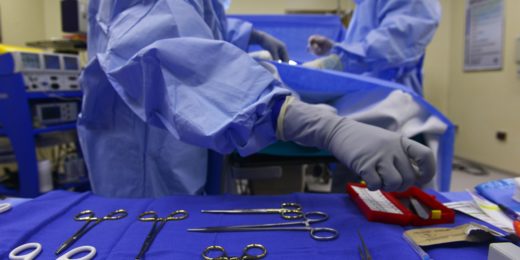
Published by
Stanford Medicine

Category

Surgery

Global Health, Medical Education, Stanford News, Surgery

Teaching surgeons new skills for medical missions

teaching-surgeons-new-skills-for-medical-missions

Surgeons practice drilling burr holes during Stanford course

Sherry Wren, MD, a general surgeon at Stanford, has volunteered multiple times for humanitarian missions in Africa with Doctors Without Borders. There, she has treated patients for everything from head traumas to difficult births to gunshot wounds; in the process, she has learned to use hand drills for brain surgery and papaya paste as a salve for severe burns, as well as how to serve as her own anesthesiologist while operating on a patient.

This month, Wren taught a continuing medical education course at Stanford to pass along the skills she learned from these first-hand experience to other surgeons and physicians interested in volunteering for similar medical missions. She recruited experts in neurosurgery, ob/gyn, and other fields to help teach the course, and she drew a large and appreciative crowd of students.

In today’s issue of Inside Stanford Medicine, I describe the course (which she calls a “labor of love”), Wren’s “MacGyver-like skills,” and her ability to “make do” with whichever supplies are available:

Developing countries may not have well-stocked supply closets; there may be no blood bank nearby; anesthesia may be limited; sonograms may be nonexistent.

“We wanted to make physicians understand that it’s all about somehow ‘making it work,’ Wren said. ‘You survive on your wits, making do with what’s on hand.”

The course itself was something of a lesson in MacGyver-like inventiveness. Students used pigs’ feet to practice ligament repair. Breech births were simulated from sleeping bags. An orthopaedic company donated thousands of dollars worth of fake bones; hand drills ordered online were used for bone-drilling practice.

Previously: What I did this summer: Stanford medical student works to improve pediatric surgical care in Tanzania
Photo by Sherry Wren

Medical Education, Surgery, Technology, Videos

Developing a universal translator for surgeons

developing-a-universal-translator-for-surgeons

Steven Schwaitzberg, MD, chief of surgery at the Cambridge Health Alliance at the Harvard Medical School Teaching Hospital, is on the hunt for technology to allow for video conferencing and real-time translation in a wide range of languages. As he discusses in this recently posted TEDxBeaconStreet talk, he plans to use the universal translator to teach surgeons around the world to perform laparoscopic surgery.

Despite all the existing translation software currently on the market, finding a communications system that’s good enough to teach surgery has proven difficult. So Schwaitzberg is working with IBM Human Ability and Accessibility Center to string together technologies to work towards building a universal translator for surgery. He demos the prototype in the above video and explains how, in addition to training surgeons in other countries to perform minimally invasive procedures, it could create a dialogue and help physicians in the United States understand how to reduce health costs while improving outcomes. It’s worth watching.

Previously: Clementines help surgeons-in-training to practice and Surgery for chronic acid reflux goes scarless

Cardiovascular Medicine, Stanford News, Surgery

‘Snorkel’ stents create lifeline to organs in method of treating complex abdominal aortic aneurysms

snorkel-stents-create-lifeline-to-organs-in-method-of-treating-complex-abdominal-aortic-aneurysms

It’s been called the chimney technique, and it’s been called the double-barrel technique. But Jason Lee, MD, prefers to call it the snorkel technique.

In the latest issue of Inside Stanford Medicine, I write about Lee, one of the most experienced physicians in the world at using this minimally invasive procedure to treat complex abdominal aortic aneurysms. In such cases, the aneurysm, a balloon-like bulge, extends very close to or beyond one or more of the aorta’s branch arteries, such as the renal arteries. This can make it challenging to use a stent graft, a small tube, to bypass the section of weakened arterial wall without obstructing blood flow to the branch arteries.

But Lee is able to circumvent this problem by placing one or more additional stents, which when deployed look like snorkels,  adjacent to the main stent to create pathways for blood to reach branch arteries.

“The Europeans like to call this the ‘chimney graft,’ but in a chimney the smoke is going up, right?” Lee told me. “I don’t think that analogy is quite right because the blood for the kidney or visceral organ isn’t going up, like smoke, through a stent; it’s going down — like the vital air that comes down through a snorkel.”

Lee and Ronald Dalman, MD, have performed more than 60 snorkel procedures in the past three years, and my piece describes how they recently used the technique to treat Geraldine Vitullo, a 65-year-old grandmother from Visalia, Calif.

Schematic drawing of “snorkel” stents adjacent to main stent reproduced with permission from the Journal of Endovascular Therapy.

Research, Surgery

Researchers draw inspiration from mussels to create new bioadhesive for surgery

More and more often researchers are turning to nature to develop new medical tools. Around the globe teams are studying porcupine quills to design adhesives that can bind internal tissues more securely, spider webs to create strong, easily removeable bandages, beetle wings to develop ultra-sensitive electronic skin and mosquito mouths to invent pain-free needles.

Now a group of researchers is working to create a new non-toxic bioadhesive that works well in wet environments based on the chemistry that allows mussels to stick to underwater surfaces. Medical News Today reports:

In recent decades bioahesives, tissue sealants and hemostatic agents became the favored products to control bleeding and promote tissue healing after surgery. However, many of them have side effects or other problems, including an inability to perform well on wet tissue.

“To solve this medical problem, we looked at nature,” said Jian Yang, associate professor of bioengineering at Penn State. “There are sea creatures, like the mussel, that can stick on rocks and on ships in the ocean. They can hold on tightly without getting flushed away by the waves because the mussel can make a very powerful adhesive protein. We looked at the chemical structure of that kind of adhesive protein.”

Yang, along with University of Texas-Arlington researchers Mohammadreza Mehdizadeh, Hong Weng, Dipendra Gyawali and Liping Tang, took the biological information and developed a wholly synthetic family of adhesives. They incorporated the chemical structure from the mussel’s adhesive protein into the design of an injectable synthetic polymer. The bioahesives, called iCMBAs, adhere well in wet environments, have controlled degradability, improved biocompatibility and lower manufacturing costs, putting them a step above current products such as fibrin glue and cyanoacrylate adhesives.

Previously: Porcupine’s quills inspire new types of adhesives, needles, Researchers turn to spider webs to design improved medical tape, Researchers look to gecko’s ultra-sticky feet to improve adhesion of bandages, sutures when wet, Beetle wing design inspires ultra-sensitive electronic skin and Researchers turn to mosquito to design painless needle
Photo by U. S. Fish and Wildlife Service – Northeast Region

Stanford News, Surgery, Videos

How orthopedic surgery straightened out one man’s life

how-orthopedic-surgery-straightened-out-one-mans-life

Scoliosis is a painful and debilitating condition that causes the spine of an individual to curve like an “S.” For Jerry Stark, having degenerative scoliosis meant he could no longer work or even live much of a life: He was so bent over that he couldn’t see himself in his bathroom mirror, and his pain grew so great that life became all about managing the pain and little else. Stark eventually came to see Stanford orthopaedic surgeon Ivan Cheng, MD, who performed a minimally invasive technique that changed his life.

Watch the video to meet the new Jerry Stark.

From Dec. 24 to Jan. 7, Scope will be on a limited holiday publishing schedule. During that time, it may also take longer than usual for comments to be approved.

In the News, Research, Surgery

Porcupine’s quills inspire new types of adhesives, needles

porcupines-quills-inspire-new-types-of-adhesives-needles

Drawing inspiration from the mechanical features of porcupine quills, researchers at MIT and Brigham and Women’s Hospital in Boston are developing less-painful needles for injections and adhesives that can bind internal tissues more securely.

Researchers studied the North American porcupine, which has about 30,000 barbed quills measuring several centimeters long. The tip of each quill is covered in microscopic barbs that allow the quills to penetrate tissue with very little force and make them quick difficult to remove. After determining how the quills achieve this unique combination, researchers designed plastic quill replicas that closely mimic the penetration force and gripping power of natural quills. Their work is described in a paper (subscription required) published yesterday in the Proceedings of the National Academy of Sciences.

Michael Longaker, MD, a pediatric plastic and reconstructive surgeon at Stanford, commented on the findings in an MIT release:

The discovery that barbed quills require so little force to penetrate skin is both unexpected and fascinating, says [Longaker]. Devices that mimic the quills could be very helpful in securing tissue during and after surgery, he says. “That combination of ‘easy to get in’ and ‘difficult to pull out’ would be really appealing,” says Longaker, who was not part of the research team

Researchers are now working on developing quill-inspired adhesives from biodegradable materials.

Previously: Researchers turn to spider webs to design improved medical tape, Researchers look to gecko’s ultra-sticky feet to improve adhesion of bandages, sutures when wet and Beetle wing design inspires ultra-sensitive electronic skin
Photo by Wagner T. Cassimiro

Patient Care, Research, Surgery

How music therapy may benefit surgery patients

Patients scheduled for a trip to the operating room may want to update their pre-procedure checklist to include “create surgery playlist.” A review study (subscription required) conducted by researchers at the University of Kentucky offers more evidence that listening to music can reduce patients’ pain and aid in recovery.

In examining the use of music before, during and after surgery, researchers found that listening to tunes during all three stages proved beneficial. Overall, patients who listened to music were less anxious, required less sedative medication, recovered more quickly and reported better satisfaction with their medical experience. But while some studies show that listening to classical music could yield the most positive results, the latest findings underscore the importance of taking into account patients’ musical tastes. Psych Central reports:

The researchers say that music selected by trained personnel is preferred because specific guidelines should be followed to maximize the positive effect on patients, however the patient’s musical tastes should still be considered.

The researchers suggest that several playlists be offered so the patient can choose the one that best suits their tastes.

The researchers also note that the tempo, rhythm and volume of the music should be carefully controlled to maximize the positive effect. Calm, slow, gentle music was shown to produce the most positive results and facilitate relaxation and pain reduction in patients, they said.

Previously: The anxiety and pain reducing powers of Adele, Can music benefit cancer patients? and Prescription playlists for treating pain and depression?
Photo by William Brawley

Cardiovascular Medicine, Health Costs, Research, Stanford News, Surgery

Is stenting or surgery better for diabetics? New study provides answer

is-stenting-or-surgery-better-for-diabetics-new-study-provides-answer

You may have heard about the new research showing that bypass surgery is better than stents for diabetics: In a Mount Sinai School of Medicine study of 1,900 diabetics with multi-vessel coronary disease, heart bypass surgery increased the five-year survival rate by 30 percent compared to the use of artery-widening stents.

I spoke with Mark Hlatky, MD, professor of health research and policy and of cardiovascular medicine at Stanford, who said that this study should settle the 17-year debate on bypass-vs-stent effectiveness with “compelling evidence.”

In a New England Journal of Medicine editorial, Hlatky goes on to say that many of the stent procedures today are performed on the fly, during a diagnostic angiogram, with the same physician making the diagnosis, recommending the treatment, and performing the procedure. He feels that these new findings will result in a change for the better in clinical practice and will enable patients to be better informed about their choices.

This study should settle the 17-year debate on bypass-vs-stent effectiveness with ‘compelling evidence.’

“Patients and their doctors need to allow time for discussions on which procedure should be done, based on outcomes that are important to them,” said Hlatky. “They need time to digest the information, discuss it with family members and members of the heart team, and come to an informed decision.”

It’s worth noting that cost may be a factor in treatment discussions. Bypass surgery costs more than implanting stents, but because it results in fewer deaths and heart attacks, it is worth the front-end expense, said researchers on the cost-analysis part of the study. After factoring in the stent-related costs of new heart attacks and follow-on operations to re-open arteries, bypass surgery still costs about $3,600 more than the stent procedure over five years.

Previously: New test for heart disease associated with higher rates of procedures, increased spending and To stent or not to stent: not always an easy answer

Image of the Week, Surgery

Image of the Week: Skin cake

Last weekend, specialty bakers and pathologists from across the United Kingdom joined forces for a disease- and anatomy-themed cake festival at  St Bartholomew’s Pathology Museum at Queen Mary, University of London. The event, titled Eat Your Heart Out, included a bake sale and lecture series.

This image shows Gillian Bell’s skin cake, a rich fruit cake covered in stitched surgical marzipan. More images of the ghoulish goodies can be viewed on the museum’s Tumblr.

Via Shots

Orthopedics, Stanford News, Surgery

Examining the impact of psychological distress on soldiers’ spinal injuries

The latest issue of The Spine Journal is devoted to casualties of war and includes some interesting insight into how the emotional stress of combat can impact soldiers’ spine injuries.

A key finding reported on in the issue is that non-battle spinal pain is a leading cause of medical evacuation for those deployed in Iraq and Afghanistan. Among other noteworthy findings, as outlined in a journal release:

  • There have been 10 times as many long-term spinal-pain casualties unrelated to battle as blast injuries
  • 60 percent of veterans seeking care for spine problems have serious continued psychological distress
  • Less than one percent of military-aged Americans serve in the military (compared to nearly 50 percent during World War II), which means that veterans’ burdens after combat are now far less visible to the general public

In the release, Stanford orthopedic surgeon and journal editor-in-chief Eugene Carragee, MD, who wrote several pieces on spinal injuries for this issue and has done two tours of duty in Iraq himself, comments:

While some of the findings in this special issue are new and specific to these long wars—such as the devastating effects of IED blasts—it’s clear that we need to re-learn some very old lessons about war… We repeatedly send young people into combat to experience the worst psychological stressors possible, push them beyond endurance, and yet on the home front there is little to no collective understanding of the wars they fought or the experiences of deployment and combat. Veterans seeking care for spine problems at home are shown to have continued serious psychological distress, associated with exposure to combat. This is a serious public health issue that will continue for many years.

Previously: Stanford and other medical schools to increase training and research for PTSD, combat injuriesAs soldiers return home, demand for psychologists with military experience grows, Helping brain-injured soldiers return – safely – to the battlefield and Are veterans with PTSD at higher risk for medical illnesses?
Photo by Staff Sgt. Stacey Haga/U.S. Air Force

Stanford Medicine Resources: