Stem cells are increasingly making the leap from lab to clinic, and therapies that use them have the potential to transform patients’ lives with dramatic treatments and cures.
But reaping stem cells’ full benefits requires a detailed understanding of the complex relationships between the cells and their environments -- whether in a lab dish or a patient’s body. That was an important takeaway message from the second annual symposium of Stanford’s Center for Definitive and Curative Medicine, which I attended last week. At the one-day symposium, ten Stanford scientists presented the results of their latest work to develop cell- and gene editing-based therapies and deliver them to patients. The day also included two keynote presentations by distinguished scientists from other institutions, as well as a talk by a local father who is helping to change the way rare diseases are studied.
“What you saw today is what the Center for Definitive and Curative Medicine is now, and what we can become,” said Maria Grazia Roncarolo, MD, the center’s director, in her remarks at the conclusion of the day. The center has 20 therapies based on stem cell biology and gene editing in its pipeline, Roncarolo told the audience. “The CDCM wants to be a home for every Stanford scientist who wants to translate fundamental discoveries in stem cell biology and gene editing into new therapies,” she added.
One big challenge, where stem cell therapies are concerned, is that these living cells have complex interactions with their environments. Several scientists presented data that illustrated this idea in different ways. For example:
- Sloan Kettering’s Michel Sadelain, MD, PhD, a pioneer of using engineered immune cells called CAR-T cells to treat cancers such as leukemia, described his team’s efforts to understand a potentially deadly side effect of this cell-based treatment. Called cytokine release syndrome, it is characterized by inflammation so severe it can endanger the patient’s life. So far, in mouse studies, Sadelain’s team has learned that the storm of problematic inflammatory molecules comes not just from the engineered immune cells but also from the animal’s own immune cells. “Multicellular interaction causes CRS, not just CAR-T cells,” Sadelain said. Now that they know this, his team thinks it may be possible to design safer CAR-T cells that don't trigger the complication. They’ve also found that CRS is less likely to occur in patients who have fewer cancer cells to start with, evidence that it may make sense to start CAR-T therapy earlier in the course of disease.
- In a basic-science talk, Stanford developmental biologist Roeland Nusse, PhD, described his work to identify liver stem cells. “It’s a general paradigm in stem cell biology that blood vessels are an important source of signals for maintaining stem cells,” he said. “The blood vessel is the source of the stem-cell-renewing signals.” He told how his team has shown that liver cells located near the liver’s central vein have “stemness,” or cell behaviors characteristic of their self-renewing properties – and the double-edged quality this confers. Not only can the cells help repair and regenerate the liver, they also give rise to liver cancer.
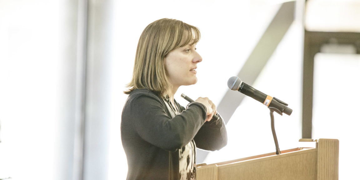
- Materials science engineer Sarah Heilshorn, PhD, is studying how to grow stem cells in a highly controlled way in large quantities, a problem that must be solved before cell-based therapies can be made widely available. Her lab is developing protein-based 3-D gels that could be used as a growth matrix for the cells. “Real stem cells live in three dimensions, not two dimensions on a dish,” she said. The cells need to be able to remodel the gel environment, her team has found. “As cells secrete enzymes, they make tunnels in the gel that let them touch their neighbors,” she explained. “They need remodeling time to form handshakes. That then allows appropriate differentiation when we hit them with differentiation cues.”
- Surgeon Michael Longaker, MD, is examining bone healing in diabetes. It’s long been known that fractures heal more slowly in people with diabetes than those without, but no one has understood why bone-regenerating stem cells work so much more slowly in the diabetic environment. Longaker’s team is demonstrating in diabetic mice that adding one specific cell-signaling protein to the fracture site appears to solve the problem. “I can see a pathway from fundamental to clinical going forward,” he said.
It will be exciting to see how specialists from different disciplines continue to bring their expertise to solving the big challenges in stem cell and gene therapies.
Photo of Sarah Heilshorn explaining how cells form handshakes by Timothy Archibald