Ask any student what they learned in their early biology curriculum and they’re almost guaranteed to say, as if by reflex, “the mitochondrion is the powerhouse of the cell.” It’s true, mitochondria play a crucial role in the body’s energy production: taking in cell nutrients, breaking them down, and creating molecules that fuel the cell.
Our understanding of the way mitochondria behave, however, remains incomplete, because methods of assessing their function has required either removing them, thus disrupting cell structure and signaling pathways, or using a technique in intact cells that relies on averaging cell populations, eliminating the ability to distinguish cell-to-cell heterogeneity and risking the loss of information.
Until now, that is.
During a recent Child Health Research Institute talk by pediatric cardiologist Daniel Bernstein, MD, I learned the importance of taking a closer look at mitochondria. He discussed an imaging program he and colleagues use to look at the differences between individual mitochondrion to better understand their behavior within cells.
Scientist Giovanni Fajardo, MD, who works in the Bernstein lab, explained that the computer program, in conjunction with fluorescent tagging, allows the researchers to examine mitochondrial function in individual live mouse cardiac cells, as well as in individual mitochondrion within a single heart cell (where mitochondria make up about a third of the cell's volume by weight, Bernstein said), in real-time.
Bernstein explained that the imaging method allows them to see “small, temporal, but important changes in mitochondrial dynamics during stress [chemically induced to simulate exercise or cardiac injury] that aren’t shown in the standard approach.”
The lab’s imaging technique shows that even in normal cardiac cell populations, where uniformity in shape and function has long been assumed, not all cardiac mitochondria produce the same response to stress stimuli. Now, they are working to understand how this heterogeneity affects cardiac conditions and diseases where dysfunctional mitochondria play a key role, such as Parkinson’s disease.
Bernstein concluded with a powerful takeaway:
The world is much more heterogeneous than you may think. The techniques being applied to understand cell behavior in the past 30 years ignore a lot of those differences. As people begin to think about how a drug affects a certain biological process, understand it may affect only, say, 20 percent of the cells and not the rest, and that there can be some useful hidden information on why some cells are affected and not others.
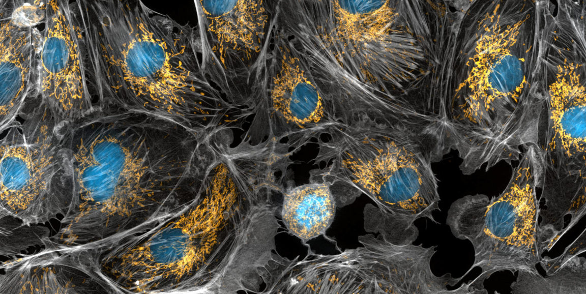
Photo of cells with mitochondria in yellow by Torsten Wittmann, University of California, San Francisco