A study in Nature details a discovery with potential clinical significance for treating eating disorders such as anorexia.
To make that discovery, Stanford researchers had to develop a "first-time-ever" way of teasing apart two separate but closely intertwined sets of identical-appearing neurons in the brain.
The study's senior author — bioengineer, psychiatrist and inventor Karl Deisseroth, MD, PhD — is heralded as a pioneering developer of optogenetics, a technology for subtly modifying selected nerve cells, or neurons, within an awake, freely moving animal's brain so those neurons's signaling can be turned "on" or "off" at the flip of an external switch. Investigators can then watch and see if, and how, the animal's behavior changes in response. This, in turn, lets them map the functions of ever larger, more widely distributed and heterogeneous circuits in the brain.
Two common behaviors about whose underlying neuronal circuitry virtually nothing is known are, in science-speak, "feeding-associated" and "social-interaction" activities. To the rest of us, this translates to eating, drinking, and being merry — or, as the case may be when presented with food in a social context, intimidated.
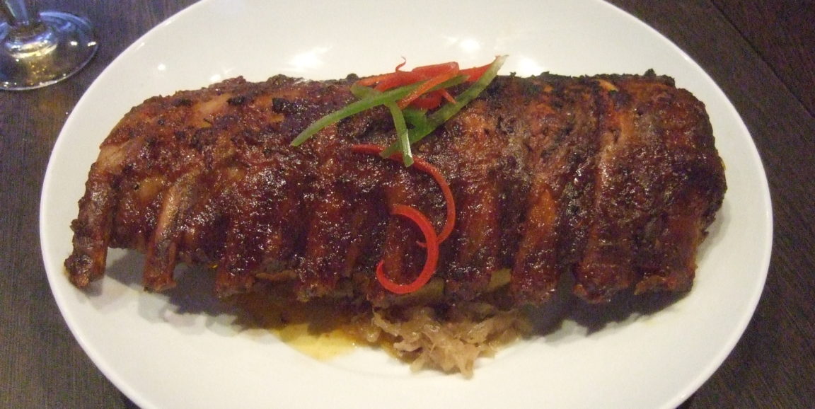
“We know social situations can inhibit the urge to eat,” Deisseroth told me when I interviewed him for a news release about the study. "You’re not going to dive into that plate of ribs when you’re dining in the presence of royalty.”
To get to the neurological bottom of the behavioral conflict, Deisseroth's team focused on a part of the mouse brain called the orbitofrontal cortex, a sheet of cells that, in both mice and humans, lies on the brain’s outer surface toward the front of the organ. This brain region, which is similar in the two species, has been shown in human imaging studies to be active when subjects are wishing for, seeking, obtaining and consuming food, or when they’re socially engaged.
But exploring the interactions of "feeding" and "social" orbitofrontal-cortex circuits was guaranteed to be tricky. From my release:
'It's not as if there is a cluster of ‘feeding’ neurons and another cluster of ‘social’ neurons sitting in two neatly labeled clumps in the orbitofrontal cortex, so you can just position an electrode in one or the other cluster and find out all you need to know,' Deisseroth said. The neurons driving and responding to these different activities are interspersed, scarce and scattered throughout the orbitofrontal cortex like sprinkles on a cupcake. Plus, they all look pretty much the same.
So the investigators designed a sophisticated system for simultaneously stimulating and monitoring activity in multiple designated neurons. They could determine which orbitofrontal-cortex neurons were active during mice's feeding-associated or social activities, or both, or neither. Then they could optogenetically stimulate numerous neurons dedicated to one or the other activity and watch what happened.
They found that stimulating a mere handful of "social-responsive" neurons induced a prompt drop in mice's food-seeking behavior.
Now that they know which neurons actually matter for which behavior, the researchers can examine them more closely for any differences that distinguish the two kinds from one another — which could lead to new drugs that reduce social inhibition of food consumption among people with anorexia.
Photo by Yun Huang Yong


