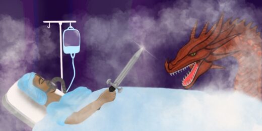
About 10 years ago, I wrote an article for our magazine, Stanford Medicine, about a remarkable new method scientists in the lab of Stanford hematologist Calvin Kuo, MD, PhD, had developed for culturing intestinal tissue in the form of little hollow spheres.
I called these mini-organs, which can be up to a quarter-inch in diameter, "gutballs." (Researchers are calling them "enteroids" these days.)
The little fellows even contract, I wrote, just like the real McCoy:
The intestine is, in its essence, a tube surrounded by rings of had smooth muscle that periodically undergo rhythmic, sequential contractions followed by relaxations. This squeezes food through the tube -- a process called peristalsis. Along the way, the food gradually gets absorbed. Getting anything even remotely similar to happen in a petri dish... hasn't been easy. In fact, until just a few years ago it had never been done, although people have been trying for more than 30 years.
Enteroids' research potential was immense. Armed with a microscope, for example, you could observe how different bacterial pathogens infect gut tissue. You could pour a drug into the dish and monitor its antibacterial activity, and its side effects. You could alter a single gene in one of the several cells types of which intestinal tissue is composed and look inside the dish to watch how that affects peristalsis, or you could add a substance to the culture medium and see whether it causes cancer.
But there was a problem. The real action in a working intestine takes place on the inside of the tube, through which food flows, good or bad bacteria gain a toehold, and ingested carcinogenic substances occasionally kick-start tumor development.
The inside-facing and outside-facing surfaces of the cells comprising the main layer of tissue in both enteroids and real intestinal tissue differ substantially. They absorb things differently, their secretions and excretions differ, they host different surface molecules.
So it's really the insides of enteroids that are of interest to scientists. But it's the outsides of the enteroids that actually get exposed to the drugs in the dish, and that are actually visible to microscopists -- it's enough to give professors and postdocs alike conniptions.
Why not flip them inside out? That, as you can imagine, is easier said than done, but as you can see in this little time-lapse movie, researchers have done it.
The movie's left panel shows an enteroid chilling out while it's embedded in a hydrogel scaffold that's initially required to generate and stabilize enteroids. The right panel depicts an enteroid flipping inside-out once the scaffold is removed -- a happy outcome after a lot of work by Stanford microbiologists Manuel Amieva, MD, and Denise Monack, PhD, and their shared postdoc Julia Co, PhD, who teamed up with Kuo's lab to figure out the details of how to turn those gutballs inside out.
Their successful efforts are described in a paper published in Cell Reports.
Now they can monitor drug effects on the inner gut lining just by pouring the drug into the culture medium, or zero in on specific molecules on the (formerly inner) surfaces of cells composing that lining as they get latched onto by different types of invading bacterial pathogens. Their resulting observation that two prominent intestinal pathogens, salmonella or listeria, have quite distinct ways of doing so is a step toward finding more effective ways of dealing with these infectious microorganisms.
Photo by Raphaël Biscaldi; Video by Mar Margalef Catala, PhD