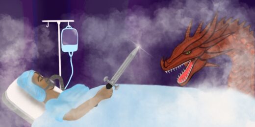
For many years, physicians believed they had less than six hours after a stroke began to prevent a patient's brain cells from dying and to save the patient from death or disability.
But, from the beginning of his career, Gregory Albers, MD, professor of neurology and neurosurgery, questioned this conventional wisdom. Over the course of three decades, he and a team of Stanford Medicine researchers tested and perfected a new imaging technique that would ultimately expand the window of time for safely restoring blood flow in a stroke patient's brain.
In "Opening stroke's window," a story in the latest issue of Stanford Medicine magazine, Albers' journey to help treat more stroke patients unfurls, highlighting how just a few hours can make a big difference to patients' lives.
"It was a huge challenge," Albers said of his quest to improve outcomes for stroke patients. "We were dealing with the No. 1 cause of disability worldwide, and there was no treatment."
When Albers began his research, it took physicians hours to get images of a stroke patient's brain -- and by then the damage had already been done. He and his team discovered an alternate way to see which brain cells had been compromised and identify if any tissue could still be saved if physicians intervened.
Science writer Amy Jeter Hansen, the author of the story, explains:
When brain cells begin to die during a stroke, they lose the ability to maintain a balance of water inside and outside the cell. Conventional techniques could capture images of dead cells swamped with water hours later, but [physicist Michael Moseley, PhD,] wanted an earlier indication of what was happening.
In a late-night laboratory insight, he discovered that he could track a stroke's damage to brain tissue as it happened through an MRI technique that was more sensitive to the movement of water in cells. Scans created through diffusion-weighted imaging showed a clear difference between cells that had no active transport of water and those with normal water-shuttling activities.
In short, Moseley had found a way to watch brain cells die in real time.
As the team rounded out their initial tests of the imaging, the data suggested that about half of stroke patients could still benefit if the clot blocking their cerebral artery was removed up to 12 hours after stroke symptoms began. Of course, there was more work to be done. Albers and his team continued with a series of trials, and in 2016, began a clinical trial that used the technique to guide treatment decisions.
The results were definitive. As Hansen writes in the story:
The data showed that the imaging technique Albers and his colleagues had refined over so many years could help physicians determine when they could do more for a stroke patient and substantially improve the patient's recovery.
Three months after a stroke, 45% of the patients who received a thrombectomy six to 16 hours after their first symptom were functionally independent, compared with 17% who received standard care. Among patients receiving the thrombectomy, 14% died within three months of having a stroke, compared with 26% in the control group.
It was the validation Albers needed. And he knew it would spark a widespread change. "This is going to change the world," he thought at the time.
He was right.
Read the full story about Albers' quest to bring better treatments to stroke patients here.
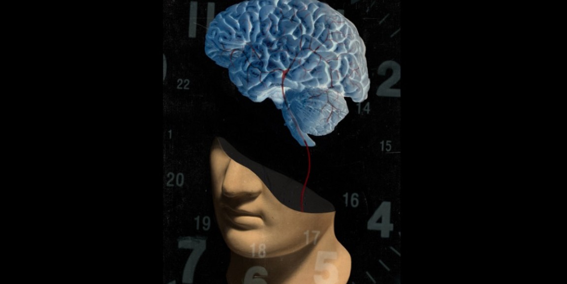
Illustration by Jonathon Rosen