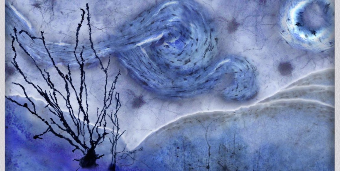
Shades of blue swirled across a dark night sky recall a tantalizingly familiar image. Amalia Perna, postdoctoral scholar at Stanford Medicine calls it "The Van Golgi," an ethereal rendition of Vincent van Gogh's "The Starry Night" that trades a canvas and paints for mouse brain sections and the Golgi-staining technique, which helps scientists image neurons.
One of 57 submissions, Perna's rendition of the masterpiece was awarded the Grand Prize in a recent scientific image competition hosted by the Wu Tsai Neurosciences Institute. The Andrew Olson Scientific Image Awards competition recognizes the creativity and expertise of the imaging community at Stanford, providing scientists an opportunity to demonstrate the more artistic side of their research.
Perna works in the lab of pathology professor Thomas Montine, MD, PhD, investigating neurodegeneration prevention and recovery. She explores the molecular mechanisms of brain cells following exposure to toxic stimuli or disease. Much of the data Perna analyzes comes from mouse models, which also served as building blocks for her artwork.
I spoke to Perna to learn more about her artwork and how she views the intersection of art and science.
What was the inspiration for 'The Van Golgi'?
I started thinking about how the Golgi-stained sections I imaged in the previous weeks could be seen as art, even more strikingly than they already appear under the microscope. My original idea was to reproduce an old piece of art. It may have been the harmony between the bright blue color of the staining chemicals and the intense black from the metal reaction that pointed me to van Gogh's "The Starry Night." The first resemblance I noticed was between the fascicular nature, a sort of clustered swirl, of the corpus callosum, a thick nerve tract that connects the two brain hemispheres, and the style that van Gogh used to paint the swirling clouds.
How did you build on the science to make the image?
Everything fell into place. I started noticing that the rounded neurons of the hypothalamus, a region of the brain that helps control things like body temperature, thirst and other aspects of bodily maintenance, could be used for the stars, while the organization of the cortical neurons looked like the little village at the bottom of the hills. Pyramidal neurons of the hippocampus were perfect for portraying cypress trees in the foreground, and the perimeter of the coronal section -- characterized by a slightly darker stain -- naturally resembled the rolling hills.
I mostly used Photoshop to size, contour and stitch together images of the structures from the brain sections. I tried to find the overlap between my images and "The Starry Night" by playing with proportions and shapes while preserving as much as possible the scientific image.
What are your thoughts on the contest's goal of combining art and science?
Often, art and science are thought of as two independent worlds, with two different languages. But I see a lot of art in science, and vice versa. This contest gave me the chance to observe different expressions of beauty in the scientific world. The great variety of acquisition methods employed to get those pictures, the different species portrayed, and techniques and chemistry used are just a few examples of how much art is hidden in what scientists do daily.
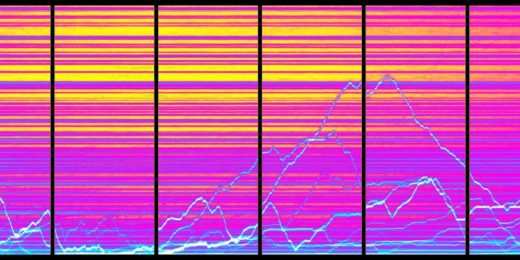
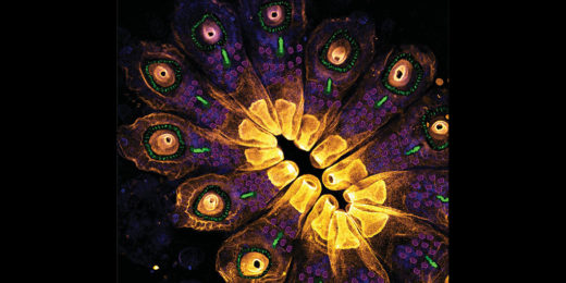
"Connected Consciousness in an Undersea Egalimind" and "A Californian Sunset" were awarded second and third place, respectively.
Image by Amalia Perna