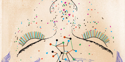
My latest story for Stanford Medicine magazine focused on something called cryogenic electron microscopy. Wait! Don't leave. It's interesting, I promise.
Think wiggly bits of SARS-CoV-2, scientists tampering with LSD to change how it impacts the brain and figuring out how to terminate HIV once and for all. Behind myriad drugs, nuanced details of disease manifestation, and so many other aspects of understanding biology, is an effort to piece together a picture -- sometimes a movie -- of how molecules look and work. That lets scientists in on all the secrets molecules hold, how they operate to perform tasks in the body and how those operations may falter during disease.
In this story, imaging is a shining beacon of knowledge, and scientists are increasingly turning to a technique that's mid-boom, called cryo-EM, to decipher biology. This technique compiles thousands of different images of the same molecule, contorted and frozen in various shapes, to build a wholistic picture of its structure.
Changes in a molecule's structure often underpin its actions. But a picture, even the clearest ones, don't capture the dynamic nature of molecules, which are critical to understanding what proteins do and how they do it. That's where cryo-EM comes in: It has the ability to detect and track proteins as they would move in the wild.
If you were an alien...
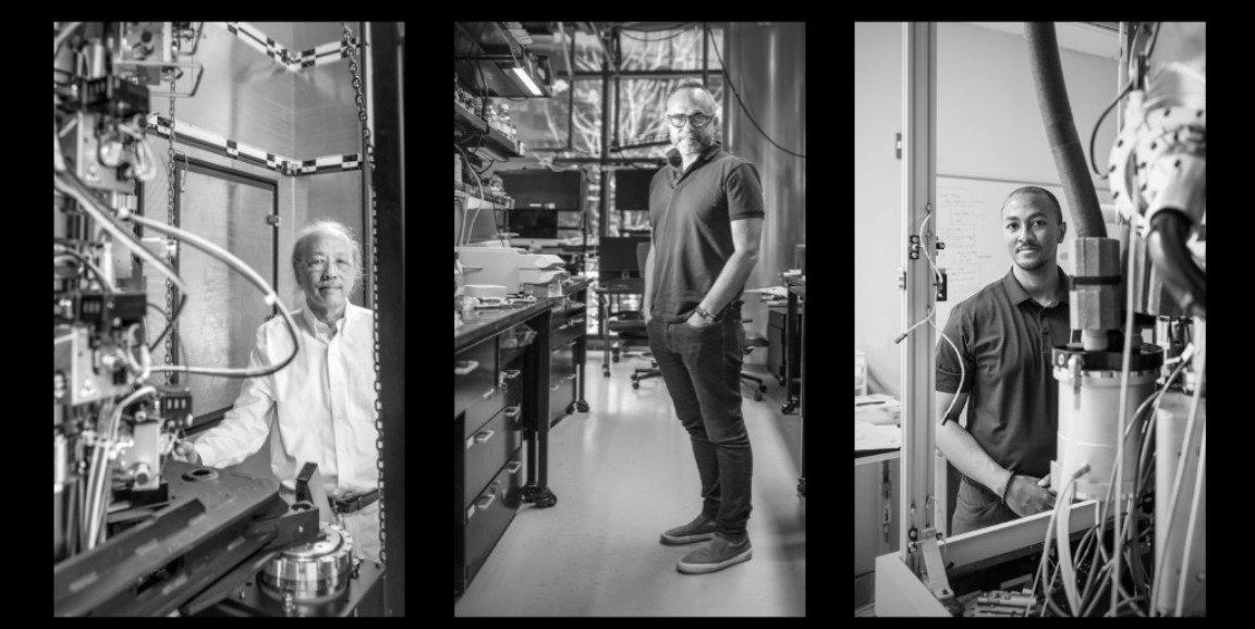
Cryo-EM expert Georgios Skiniotis, PhD, professor of molecular and cellular physiology and of structural biology at Stanford Medicine, puts it another way: Imagine you're an alien visiting Earth for the first time. Your mission: Capture the complexities of human movement through photographic evidence and return home. What might you shoot? Maybe one photo would show someone standing up; another, someone sitting down; and another, someone mid-stride.
The static images lend an idea of what a person is like -- four gangly appendages with a knob of sorts that sits up top -- all in different conformations. But the movements in between those poses are lost, as is information about function and how that person interacts with its environment. If you really want to know how humans flow from one position to another, you need something more sophisticated, with thousands of photos that are closely linked; something more akin to movie. That's what cryo-EM offers.
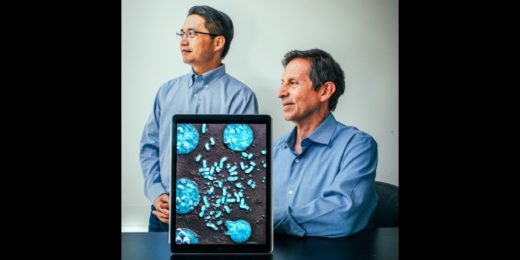
With a movie, you can see how one state transitions to another -- a premise that Wah Chiu, PhD, a professor of microbiology and immunology and of bioengineering, and Christopher Barnes, PhD, assistant professor of biology, are capitalizing on to understand how viruses, such as SARS-CoV-2 and HIV, infect cells and how to stop them.
Find out more about how these scientists are harnessing this powerful imaging technique to unearth secrets of the molecular variety to develop treatments and vaccines, and uncover what makes a molecule tick.
Photo by Timothy Archibald