Cancer drugs often don't work inside the brain. That's because a tightly structured matrix of tissue and blood vessels, called the blood-brain barrier, blocks the entry of most foreign molecules.
Figuring out which drugs can cross the blood-brain barrier -- and whether they stay active long enough to fight cancer -- typically requires harvesting brain tissue, grinding it up and using molecular analysis techniques to measure drug activity at different time points. It's an expensive and time-consuming process. But a team led by researchers at Stanford Medicine has engineered a more elegant solution: a bioluminescent indicator that makes brain tissue glow when a certain type of cancer drug is active.
"One of the great things about our method is that it doesn't require any kind of expensive imaging equipment," said Michael Lin, MD, PhD, associate professor of neurobiology and bioengineering, whose lab led the research. "One of our imaging machines was literally a black wooden box with a circular hole cut in the top, where we put a regular camera."
Using their new method, described last month in ACS Central Science, the researchers were able to screen possible cancer drugs for activity in the brain. They also identified a promising new kinase inhibitor -- a type of drug that blocks uncontrolled cell growth -- that was previously not known to cross the blood-brain barrier.
The importance of kinases
Kinases are a class of proteins that modify the activity of other proteins by adding a chemical group called a phosphate, in a process called phosphorylation. Phosphate groups act like on-off switches for many proteins, and out-of-control kinase activity causes hyperactive cell growth and excessive cell survival, contributing to a variety of cancers and neurodegenerative conditions. Drugs that block this activity, called kinase inhibitors, are a mainstay of many cancer treatments.
"Kinase inhibitors have been really successful in extending the lives of cancer patients," said Lin, referring to their ability to block the runaway cell growth that's responsible for many solid tumors. "But there's currently no kinase inhibitor approved specifically for use in brain cancers."
To investigate whether kinase inhibitors could be effective against brain cancers, the researchers sought a noninvasive way to determine which kinase inhibitors cross the blood-brain barrier.
Researchers sometimes use fluorescence, which involves shining one color of light on a part of the body and measuring a different wavelength of light reflected back as a proxy for drug activity, but fluorescence cannot be easily imaged through layers of tissue. So, researchers cannot rely on it to see deep inside the brain.
A better option, Lin said, is to use bioluminescence, a biological process that converts a chemical fuel, called a substrate, into light -- think fireflies or anglerfish.
A new type of bioluminescence
The light-generating protein in fireflies doesn't create a bright enough signal for use in brain imaging. However, a new type of synthetic protein, derived from an enzyme in deep sea shrimp, glows brightly enough to be used and seen in the brain.
By modifying the synthetic protein, called NanoLuc, the researchers were able to create an indicator that glows when a kinase inhibitor is active. They created the indicator by attaching NanoLuc to two protein fragments that bind to each other when one of them is phosphorylated by a kinase. When the protein fragments bind, they pull NanoLuc apart and prevent it from glowing.
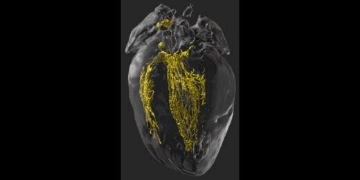
"You can think of our indicator like a Pac-man," Lin said, "where the two sides of its mouth are the pieces of NanoLuc, held apart by the kinase's phosphorylation." When a drug successfully inhibits the kinase and prevents the addition of a phosphate group, the two halves of NanoLuc join, enabling the Pac-man to gobble a chemical substrate, which is also added to the mouse's brain. The brain then glows, signaling that the kinase inhibitor has successfully passed through the blood-brain barrier and is active inside the brain.
Once the researchers created their bioluminescent indicator, they used a virus to deliver the indicator into mice, either into healthy brain cells or tumor cells, depending on the experiment. Then, the researchers treated the mice with a variety of possible kinase inhibitors to see which ones cross the blood-brain barrier effectively.
An unpredictable journey into the brain
One kinase inhibitor in particular, temuterkib, stood out as especially promising, although computer algorithms had predicted it would perform poorly.
"Understanding what makes a drug likely to penetrate the blood-brain barrier is a problem chemists have been struggling with for a long time," Lin said. Many researchers have tried to make predictions based on drug structure and size, using pattern recognition and machine learning, but have found only partial success. "It's really quite unpredictable," he added. "If it were as easy as using a computer algorithm, we would have cured brain cancer already."
Many drugs can't passively diffuse across the blood-brain barrier; they must get shuttled across by small molecule "transporters," which are specialized proteins embedded in the lining of the blood-brain barrier that can bind to drug molecules and ferry them across. Drugs must also avoid getting kicked back out by another set of proteins, which work to pump out foreign molecules.
Interestingly, some of the kinase inhibitors crossed the blood-brain barrier when tested in mice with tumors, but not in mice with healthy brains, said Yichi Su, PhD, postdoctoral scholar in neurobiology and co-corresponding author on the paper. "This shows how important it is to choose the right type of animal model when developing new treatments," he said.
Because cancer can damage the cells that make up the blood-brain barrier, the barrier may be "leaky" in the center of a tumor but intact at the edges. To make sure a drug can reach all the cancer cells, even those at the margins of a tumor, the researchers determined it's better to test the drug on mice with a fully functioning blood-brain barrier.
Making drug development accessible
Developing a good drug for the brain typically requires a costly investment in specialized lab mice and imaging equipment, making those studies infeasible for most academic labs. Using bioluminescent indicators is much cheaper, Lin said, both because the equipment is inexpensive and the imaging is noninvasive, so researchers can work with the same mice for longer periods.
The scientists plan to use bioluminescent indicators not only to screen new drugs to treat brain cancer and other neurodegenerative disorders, but also to test molecular modifications that could improve drug penetration into the brain. "We hope these bioluminescent indicators will help speed up drug development," Su said, "by allowing scientists to quickly and inexpensively evaluate potential drugs."
Photo by dTosh