It’s said that the human brain is the most complicated thing in the universe. Take that with a grain of salt, because it’s the human brain that’s saying so.
But still. The human brain is one of the great mysteries in science, and it's a tough nut to crack. How can you study its development when so much of it takes place while a child is still in its mother's uterus? Even if that weren't a show-stopper, as I've written in a recent Stanford Medicine magazine article, “Brain balls”:
You can’t exactly scoop a chunk out of someone’s living brain. And dead ones don’t tell you nearly enough.
If only you could grow a brain in a bottle, you could learn a lot about what can go wrong – or for that matter, what goes right – in early brain development. That knowledge, in turn, could help medical scientists better understand autism, epilepsy, dyslexia, and many other neurological conditions that begin in the womb.
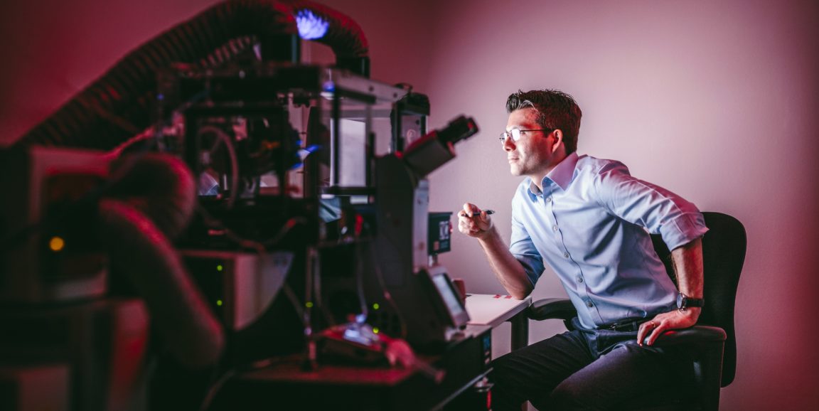
My magazine article recounts how Stanford neuroscientist Sergiu Pasca, MD, figured out how to grow brain-like blobs in lab dishes. It was no cakewalk. First he and his wife, neonatologist Anca Pasca, MD, had to get all the way from Transylvania, where they grew up (seriously!), to Stanford. Then Pasca had to overturn traditional customs for culturing nerve cells in dishes, which Anca helped Sergiu do by fiddling around with some innovations he had come up with in some of what what he calls his "Saturday experiments" at Stanford.
The eventual outcome was the sprouting, in a dish, of little balls about one-sixth of an inch in diameter, each recapitulating the architecture of the brain and replete with many (although not all) of the cell types present in a real, live human brain.
From my article:
One of the most amazing things about their brain balls was that, with not much chemical guidance, they tended to take on a default structure that’s a facsimile of the most evolutionarily advanced part of the brain: the human cerebral cortex, with all six layers you find in a living human brain.
Plus, with some chemical guidance you can get brain balls to adopt alternative structures resembling other parts of the brain. You can even hook up one brain ball resembling a brain region with a brain ball resembling another brain region and watch, real close up, how the two interact.
These tiny, signaling spheroids have given an expanding roster of researchers an astonishing front-row seat from which they can view early goings-on and goings-wrong in the human brain. Or, maybe, learn how to grow a better brain. One shouldn't rule out that possibility.
Imagine: Higher SAT scores.
Photo by Timothy Archibald