Researchers have just made the first molecular movie of one of nature’s fastest and most important biological processes — observing how retinal changes shape when hit by light.
Retinal is critical to vision and many other light-driven processes. It is a small molecule that nestles into the binding site of a specialized protein in the cell membrane. In our eyes, retinal molecules bind to opsin signaling proteins to form visual pigment molecules, which coat our retinas and help us detect light.
Scientists knew that retinal changes shape when struck by light, which activates its opsin protein to send a signal to the cell’s interior to initiate vision. But they didn’t understand the details until an international team of researchers directly visualized the molecules in motion, as recently reported in Science.
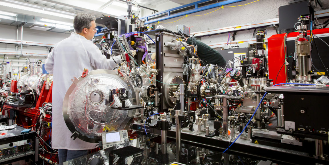
The above animation outlines the experiments they performed at the SLAC National Accelerator Laboratory.
The team created tiny crystals of retinal-protein pairs. They sent these crystals through the path of two lasers. First, they hit them with an optical laser, causing the retinal molecules to change shape. And then they hit them with ultra-fast, intense X-ray pulses, recording how the retinal and protein atoms’ locations changed over time.
They assembled these snapshots — taken over just 10 trillionths of a second — into a molecular movie that shows how retinal moves instants after its hit by light.
The researchers were surprised by what they saw. Previously, scientists thought the signaling was launched by the retinal pushing on the protein. However, the molecular movie shows the protein actually moves first, making room for the retinal to change shape. The proteins’ movements make the retinal more efficient, helping to explain why we can see down to a few photons of light, they explained.
“We always say seeing is believing in structural biology, and in this case it’s very true,” said Jörg Standfuss, PhD, a biologist at Paul Scherrer Institute and lead author of the paper, in a recent SLAC news release. “The molecular movie we made makes it so obvious what’s going on that you can immediately grasp it. This solves a very important piece of the puzzle of how retinal works that people have been wondering about.”
Retinal plays a key role in many different biological processes in people, animals and microbes, so this new understanding could have a wide-reaching impact. Standfuss explained in the release, “I really hope that we can now study the same reaction in many different systems. Now that we see for the first time how it works in one particular bacterial protein, I want to understand how it works in the human eye as well.”
Retinal is also important in optogenetics, a technique that uses genetic modification and light stimuli to precisely manipulate neurons in living tissue. Researchers are studying optogenetic treatments for vision, deafness, pain, Parkinson’s disease and many other conditions.
A better understanding of how retinal works may eventually lead to improved treatments, including optogenetic therapies.
Photo by Fabricio Sousa and animation by Andy Freeberg, courtesy of SLAC National Accelerator Laboratory and Paul Scherrer Institute


