A few years back, Stanford scientists Sergiu Pasca, MD, and Anca Pasca, MD, a husband-and-wife team who each run their own lab in the School of Medicine, began thinking about the sorts of experiments they could conduct if they had a way to study brains in a dish.
A new paper in Nature Medicine showcases the latest discoveries from those efforts: Using their lab model, they identified a type of developing brain cell that is profoundly changed by exposure to low oxygen levels. The cells are called intermediate progenitor cells of the subventricular zone, which is a region responsible for the growth of the cortex in the human brain.
The work is a first step in addressing a common form of brain injury in premature babies. Preemies don't breathe well -- their lungs aren't very mature, and neither are the breathing centers in their brains -- so it's fairly common for them to suffer hypoxic (low-oxygen) brain injuries. Better therapies are needed.
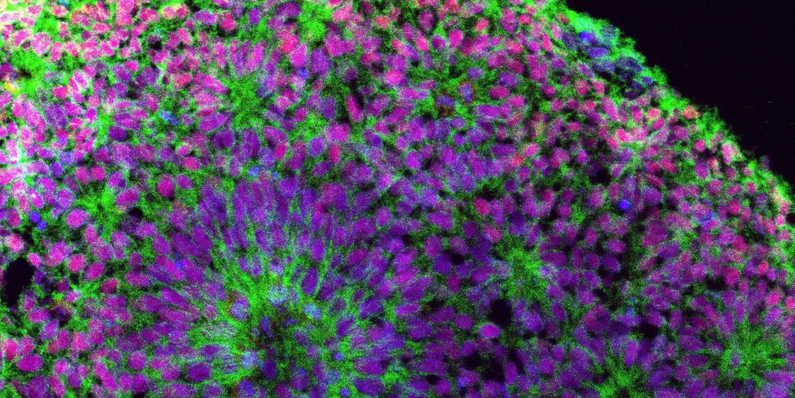
The Pascas' brain model, consisting of small, three-dimensional groups of cells grown in the lab from human-induced pluripotent stem cells, gives the researchers access to brainlike structures for conducting studies they could never do in people.
"It is quite remarkable how these blobs of cells, if provided with the right cues, will differentiate, organize and mature in a dish," Sergiu Pasca told me.
In the new study, the researchers explored the mechanism of hypoxic injury to developing brain cells. They exposed cortical spheroids, the blobs of cells, to low oxygen levels and measured resulting changes in gene expression. Our press release explains what the team learned:
They ... saw changes in a group of genes expressed in the subventricular zone, an area rich in progenitor cells that generate neurons in mid- and late pregnancy. Closer analysis showed these progenitor cells were not dying. Rather they were differentiating into neurons sooner than normal, a change could lead to fewer neurons in total in the mature brain. The pathway responsible, called the unfolded protein response, is a stress-response pathway that transitions cells from a 'growth' mode to a 'survival' mode.
'We were surprised, but it was a good surprise,' Anca Pasca said. 'Looking back, it made sense.'
Prior research showed that babies born very early tend to have thinner gray matter, with fewer neurons than normal. That's why the team thought it made sense that they observed an early shift from "growth" to "survival" in the cells that are supposed to be making lots of new neurons.
Researchers repeated the low-oxygen experiment, but added a compound to the cortical spheroids that interferes with the unfolded protein response. This time, the progenitor cells did not differentiate early. It's a clue that preemies might be helped by medications that act on the same pathway.
Image of a cortical spheroid courtesy of Yuki Miura, Pasca Lab.