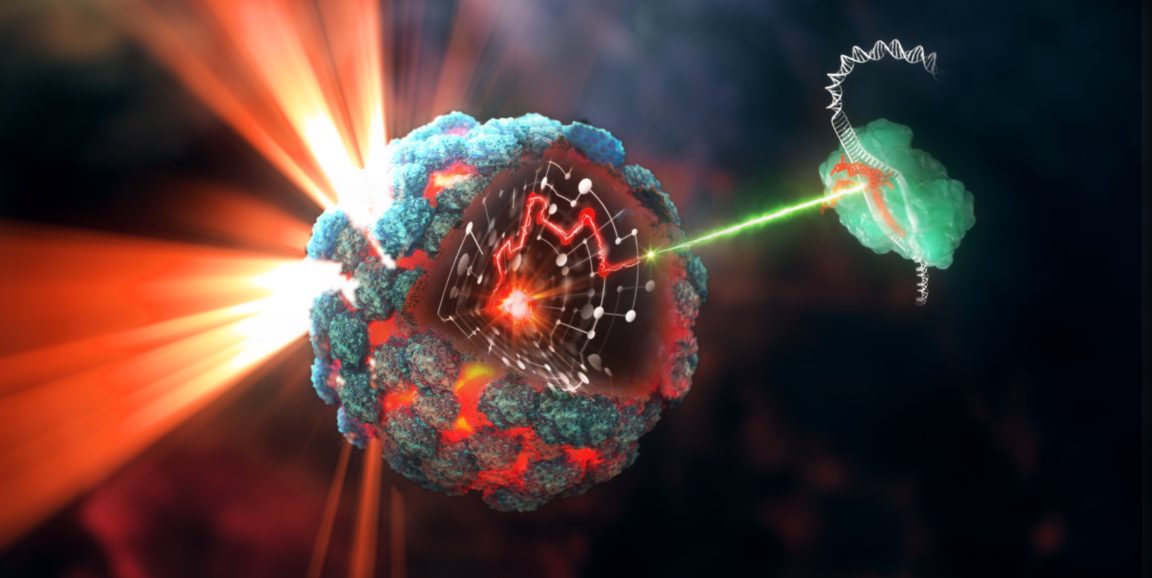
In recent months, Michael Bassik's lab members have been busy making millions of tiny round tumor "spheroids." These little round balls of cells are a type of 3D lung cancer model that Bassik, PhD, assistant professor of genetics, and his lab team are using to better understand how and why tumor tissue grows.
Lung cancer spheroids, are meant to better capture the molecular details of lung cancer found in real patients. Now with their new 3D model, Bassik and his team have discovered additional molecular mechanisms that fuel the growth of tumors.
Details of their discovery, led by postdoctoral scholar Kyuho Han, PhD, were published in Nature.
For a long time scientists investigating the molecular engine of cancer would grow tumor cells in flat layers in a dish. But that tactic has left researchers wanting.
"People have known for some time that cells in a plastic dish are not the best way to accurately model cancer in an actual patient," said Bassik. "Two dimensional models in particular don't always capture key aspects of the biology in detail."
So Bassik and his team started making more robust physical mock-ups of lung cancer tissue, which are grown from actual cancer patient cells, to better expose the drivers of tumor growth and potentially reveal clues into how it can be slowed or stopped.
Three-dimensional models of cancer are not new; scientists have been toying with building new ways to inspect cancer through 3D tissue culture for years now. But Bassik's model makes a few salient additions that set it apart.
First, these 3D spheres, which are about half the size of a grain of sand, are highly scalable, meaning Bassik and his group can make a lot of them in a relatively short amount of time -- as in millions in a few weeks. They also contain more cells than previous cultures of lung cancer spheroids, which typically comprise a few million cells. Bassik's contain at least 200 million. That's key, Bassik told me -- it allows for the final, and arguably more important, component of their model: the use of CRISPR-Cas9 to search the entire genome for new cancer drug targets.
Bassik and his team are employing CRISPR gene-editing technology to study the molecular details of these models. The idea is to use CRISPR to change the function of all genes, one per cell, to see how they affects the growth of the tumor model. That way, each spheroid is slightly different, and if its growth is slowed in a particular lung sphere, for instance, Bassik and his team know the gene altered in that model is a critical driver of lung tumor development -- and potentially a new drug target.
"We saw this happen with a gene called CPD, which seems to be required for the function of two known oncogenes," said Bassik. (An oncogene is a gene that, when mutated, can contribute to the transformation of a healthy cell to a cancer cell.) "We saw that CPD deletion prevents tumor growth in cells and also in tumors in mice."
What's more -- in the classical cells-in-a-flat-dish model, CPD's deletion had limited effect, showcasing the importance of cancer models that more realistically capture what's happening in a patient's body.
The upshot, said Bassik, is that CPD is closely tied to the successful function of a notorious oncogene, called KRAS. So far in their models, disabling CPD and KRAS kills cancer cells originally spurred by a KRAS mutation.
"Scientists haven't really considered CPD as a drug target before; in fact, it hasn't been studied much at all as a cancer target," said Bassik. "It was really only through these 3D CRISPR models that we found CPD had an effect."
Now Bassik and his team are looking to more thoroughly explore CPD as an option to fight cancer.
"We're excited to explore CPD as a drug target and to continue to develop our model to better understand what's behind cancer in patients."
Image by Jon Heras Equinox Graphics, LTD


