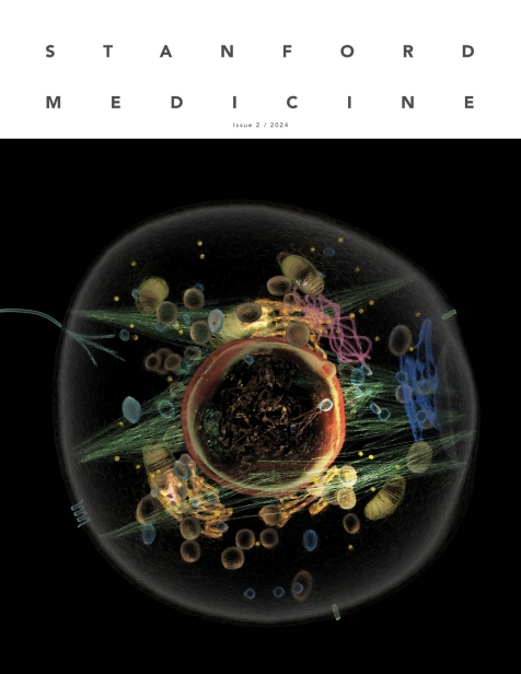
Cells may be little, but they are also fierce and powerful -- as you may have already been reminded while reading the current issue of Stanford Medicine magazine.
These tiny building blocks have a lot of the same internal structures in common, such as walls and appendages, an operation center called the nucleus, and energy reservoirs called mitochondria. What cells do with those shared structures varies so widely that new variations of cell functioning continue to be discovered.
Cells have long captivated the minds of scientists. It's why Stanford Medicine researchers were so willing to gush about their personal favorites in the magazine story headlined "My favorite cell."
Because the passion for specific cells ran so deep, we decided it was worth hearing some more cell love letters. This next group of researchers picked cells from all over the human body -- cells of all shapes, sizes and abilities. From the brain to the heart to the intestines, these cells are influential and awe-inspiring.

Laura Michele Hack, MD, PhD
Assistant professor of psychiatry and behavioral sciences
Mirror neuron
Understanding why smiles are contagious fascinated Hack when she was an undergraduate student studying neurobiology at the College of William & Mary. The neurons responsible for coaxing us to smile when we see others grinning are called mirror neurons. These cells were first described more than 30 years ago by Italian neuroscientists who showed in animals that the same neurons respond when they're picking up an object and when they're watching another animal pick up an object.
Mirror neurons get their name because they fire in the same way whether we are carrying out an action or watching someone else do it. These neurons have been found in people, through direct electrical recordings in the brains of patients undergoing presurgical planning and through experiments using magnetic resonance imaging.
"When you see someone smile, the mirror neurons in your brain that correspond to that action become activated, triggering a response that makes you feel similar emotions, albeit to a lesser degree, as if you were smiling yourself," Hack explained. "This activation also facilitates the imitation of the observed action, increasing the likelihood that you smile in return."
The exact number of mirror neurons is unknown, but they have been found all over the brain -- in networks of areas responsible for planning and executing movements, making sense of sensory information, and processing emotions. Research in people has shown that these neurons also help us understand speech and interpret the behavior of others.
Now as a psychiatrist and researcher, Hack remains mesmerized by mirror neurons -- and sees an opportunity to apply them to the study of mental illness.
"The dysfunction of mirror neurons is implicated in various psychiatric disorders, and they are thought to play a role in conditions involving impairments in recognizing emotional expressions, such as autism, schizophrenia and depression," Hack said. "I'm especially interested in how modulating activity in networks containing mirror neurons may help treat particular subtypes of depression and comorbid stress-related disorders."

Juliet Knowles, MD, PhD
Assistant professor of pediatric neurology and of pediatrics
Oligodendrocyte precursor cell
Of all the cells in the brain, neurons spend the most time in the spotlight. But neuronal function depends on a cell that works hard out of the spotlight: the oligodendrocyte precursor cell.
"These cells have a beautiful, branching morphology and have long been underestimated, but they are critical to healthy brain function and disease," said Knowles, who first encountered oligodendrocyte precursor cells during her residency at Stanford Medicine. "Each one of these cells claims a territory in our brain, and if one becomes damaged or dies, other oligodendrocyte precursor cells can replicate themselves to provide a replacement."
Oligodendrocyte precursor cells are critical to brain function because of the role they play in the production of myelin, a fatty substance that enables neurons to send and receive information via electrical signals. The axons of neurons are wrapped in layers of myelin, insulation that works like the colored plastic that sheathes electrical wire. Without myelin, the electrical signals neurons use to communicate can falter. Myelin is manufactured by oligodendrocytes, and as their name suggests, oligodendrocyte precursor cells produce oligodendrocytes.
As a postdoctoral fellow in the lab of Michelle Monje, MD, PhD, the Milan Gambhir Professor in Pediatric Neuro-Oncology, Knowles realized there might be a consequential role for oligodendrocyte precursor cells in pediatric epilepsy.
"We discovered maladaptive myelination, a process in which seizures promote altered patterns of myelination that in turn leads to further seizures," Knowles said. "My lab studies this process."
Oligodendrocyte precursor cells also contribute to changes in brain structure that happen as children develop. Their branching shape points to their role in responding to signals from other brain cells and patterns of brain activity that occur during learning. Oligodendrocyte precursor cells are also sensitive to our circadian rhythms. Knowles said understanding how these cells are altered by diseases such as multiple sclerosis, epilepsy, addiction and Alzheimer's disease could lead to new therapies that help restore myelin production in the brain.

Daria Mochly-Rosen, PhD
George D. Smith Professor in Translational Medicine
Cardiac myocyte
When the protein chemist Mochly-Rosen was in graduate school, one of her lab mates was working with muscle cells in culture. She remembers being amazed as she stared at the thin plastic dish and watched the cells rhythmically contract.
Mochly-Rosen used heart muscle cells, called cardiac myocytes, to figure out how a family of enzymes, called protein kinases C, affect the heart's beat and its response to disease.
"Early in my career, people were studying this enzyme family in cancer cells, and the enzymes were all behaving the same way, which did not make sense to me," she said. "We performed experiments on heart cells in culture, and those resulted in a breakthrough in the field. That is why cardiac myocytes have a special place in my heart."
When cardiac myocytes are outside the body and in culture, they can initially take all sorts of shapes. But they do not like being alone, and they will try to touch other cells. This touching leads to all of them eventually contracting and relaxing at the same rate. Cardiac myocytes are not like other cells in culture that will grow to such an extent that the dish becomes crowded; Mochly-Rosen said they do not like being confined because otherwise they cannot contract effectively.
In the body, cardiac myocytes are rectangular in shape and lined up in rows, like building blocks in a wall. Mochly-Rosen explained that this organization allows the cells to exert a mechanical force that has a specific direction. Cardiac myocytes lined up and beating together is what lets the chambers of the heart contract and relax, moving blood throughout the body.
"When you look at cardiac myocytes with a microscope, that allows you to look at the structure inside a cell," she said. "You realize that the contractile structures really look like springs" -- which allow the myocytes to contract and relax.
Along with neurons, cardiac myocyte cells have the highest number of mitochondria, which are like little power plants that provide energy to cells. When cardiac myocytes and other muscle cells contract, the internal springs squish the mitochondria.
"This is how the mitochondria sense how fast the heart is beating, and they can communicate that to other organs, such as our brain," Mochly-Rosen said.
The muscle cells that enable the human heart to beat are found in the hearts of other mammals, from mice to whales. A mouse heart is about as big as a fingernail, a human heart is fist-sized and a whale heart is so big a full-grown human can crawl through its chambers. Mochly-Rosen learned decades ago that these hearts are made up of cardiac myocytes that are roughly the same size -- whale hearts just have more and mice hearts have fewer than the human organ -- and she has never forgotten this fact.

Nielsen Fernandez-Becker, MD, PhD
Clinical professor of gastroenterology and hepatology
Enterocyte
It was love at second sight for Fernandez-Becker and enterocytes, cells that form the interior lining of the intestines and are involved in celiac disease and inflammatory bowel disease. She first encountered them as part of her histology rotation in medical school. When she observed endoscopic exams during her internal medicine rotation, she saw that the inside of the intestines were covered in cells with tiny, finger-like projections and that they looked like sea anemones drifting in an ocean current. She was hooked.
"The small intestinal lining is stunning, and I am still captivated by how a single epithelial cell layer can both absorb nutrients and protect us from harmful invaders," Fernandez-Becker said.
The finger-like projections Fernandez-Becker saw are called villi, and they cover the surface of the columnar enterocyte cells like bristles on a brush. The villi increase the surface area of enterocyte cells, allowing them to extract nutrients from food as it passes through the intestines. Enterocytes also make and secrete enzymes that help break down nutrients and regulate how much water is in the intestines.
"There are 20 to 30 billion enterocytes in the small intestines at any given time, and they have a remarkable regenerative capacity," Fernandez-Becker said. "They are regularly replaced, with a new layer being formed every three to five days."
The enterocytes that line the small intestine do not just nourish the body; they also form a protective barrier, acting like a bouncer for what passes through the intestinal walls. In addition, these cells interact with the immune system to help detect and neutralize pathogens.
Gastroenterologists are investigating the relationship of diet on enterocyte health and how dysfunction in these cells relates to systemic and neurodegenerative diseases.

Hawa Racine Thiam, PhD
Assistant professor of bioengineering and of microbiology and immunology
Neutrophils
When the biophysicist Thiam bought her first immunology textbook, she was disappointed. Only a couple of paragraphs touched on neutrophils, even though they account for about 50% to 75% of all white blood cells in the body.
"It was very frustrating," Thiam said. It wasn't as if everything about neutrophils was already known. "Neutrophils are the first responders to infection, but it's not really clear why or how they accomplish this."
Compared with other cells, neutrophils are lightning fast. They can move an entire cell length in one minute -- a distance that takes a cancer cell or a fibroblast a full hour to cover. And while the nuclei of most cells in the animal kingdom are round, neutrophils of different species have nuclei that are lobed, doughnut-shaped or linear. Finally, they have a repertoire of deadly force that exceeds that of other immune cells -- including extruding a net of DNA studded with antimicrobial proteins far outside their membranes to ensnare and kill bacteria.
Understanding how these quirky first responders work will inform future studies of how cells divide, specialize and function, Thiam said. "What does it mean for a cell to move?" she said. "Physically what has to happen? Membranes interact with one another; nuclei deform to let cells squeeze into tight places. This all happens very quickly and on a physical, not genetic level."
"I look at the cell as an object," Thiam said. "Geneticists look at individual genes. But it's hard for me to understand how removing one gene impacts the whole cell. It's like investigating how a car works by removing one wheel. To me, neutrophils are just super cool little objects."
Krista Conger contributed to this report.

Stanford Medicine magazine explores how the smallest units of life rule our health: The new issue of Stanford Medicine magazine covers research on cells, providing insights into basic biology, human health and the power of curiosity.


